
Medial patellofemoral ligament reconstruction is a surgical procedure indicated in patients with more severe patellar instability. The medial patellofemoral ligament is a band of tissue that extends from the femoral medial epicondyle to the superior aspect of the patella. The medial patellofemoral ligament is the major ligament that stabilizes the patella and helps in preventing patellar subluxation (partial dislocation) or dislocation. This ligament can rupture or get damaged when there is patellar lateral dislocation. Dislocation can be caused by a direct blow to the knee, twisting injury to the lower leg, strong muscle contraction, or because of congenital abnormality such as shallow or malformed joint surfaces.
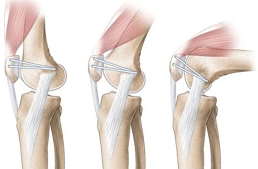
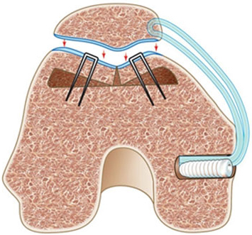
The surgical procedure of medial patellofemoral ligament reconstruction involves the following steps:
A knee brace should be used during walking in the first 3-6 weeks after surgery. Avoid climbing stairs, squatting, and stretching your leg until there is adequate healing of the reconstruction.
Rehabilitation exercises, continuous passive motion, and active exercises will be recommended.
Make an AppointmentDr. Rakesh Kumar Best Orthopedic Doctor Near Me. We strive to deliver a level of service that exceeds the expectations of our Patients. If you have any questions about our services, please do not hesitate to contact us.
Copyright © 2026 Dr. Rakesh Kumar. All Rights Reserved. Privacy Policy | Terms of Services